Asymptomatic Giant Thymolipoma Mimicking Cardiomegaly: Direct Graphy, Ct And Mri Findings
MEHMET EMİN SAKARYA, ABDUSSAMET BATUR, KEMAL ÖDEV
- Year : 2013
- Vol : 29
- No : Ek
- Page :
43-44
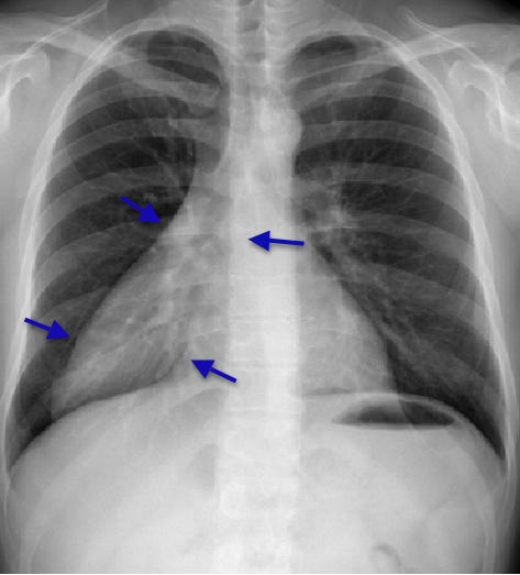
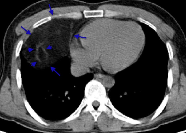
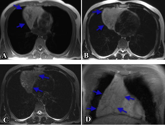
Demonstrating the imaging findings of asymptomatic giant thymolipoma mimicking cardiomegaly is aimed. A paracardiac mass was detected with 32-year-old male patient afterwards demonstration of cardiomegaly on a posterior-anterior (PA) chest x-ray examination taken for a routine control. Computed tomography (CT) showed a mass, localized in the anterior mediastinum, with smooth margins, showing no significant invasion of adjacent structures, and including fat density with fibrous component. The diagnosis was reported as thymolipoma histopathologically after transthoracic biopsy. On a preoperative magnetic resonance (MR) examination, taken for detailing the limits of the lesion, the mass showed hyperintensity on both T1 and T2 weighted images, and suppression on fat saturated T2 weighted images. No invasion of adjacent structures was detected. A thymolipoma may reach a large size without symptoms due to its great pliability. It can mimic cardiomegaly on x-ray images. Adipose tissue on CT is helpful in the diagnosis. MRI gives valuable information in determining the exact boundaries of the lesion.
Cite this Article As :
Description :
None of the authors, any product mentioned in this article,
does not have a material interest in the device or drug. Research,
not supported by any external organization.
grant full access to the primary data and, if requested by the magazine
they agree to allow the examination of data.
Asymptomatic Giant Thymolipoma Mimicking Cardiomegaly: Direct Graphy, Ct And Mri Findings
2013,
Vol.
29
(Ek)
Received : 09.07.2012,
Accepted : 09.07.2012,
Published Online : 13.08.2018
Selçuk Tıp Dergisi
ISSN:1017-6616;
E-ISSN:2149-8059;