Ct And Mri Finding Of Langerhans Cell Histiocytosis Disease
DEMET KIREŞİ, GANİME DİLEK EMLİK, ÖZLEM DÜZENLİ
- Year : 2011
- Vol : 27
- No : 2
- Page :
118-120
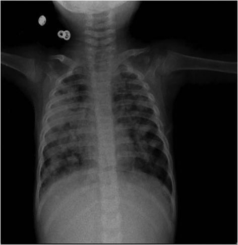
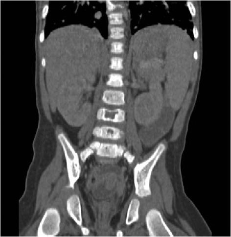
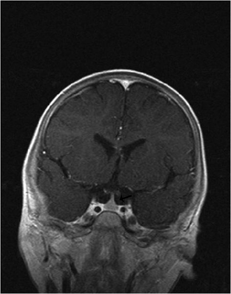
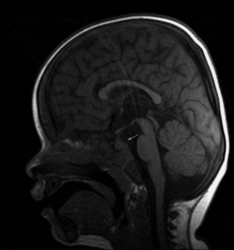
Langerhans cell histiocytosis is a group of idiopathic disorders characterized by the abnormal proliferation of specialized bone marrow-derived Langerhans cells. In this report, we present a rare case of Langerhans cell histiocytosis with CT and MRI in a 2 yearold-boy who developed symptomatic diabetes insipidus and multiple bone ,cranial, lung and spleen metastases during the disease course. A 2-year-old male patient was hospitalized due to complaints of cough, fever, vomitting, diarrhea, achromasia, weakness and rash. Chest X-ray revealed reticulonodular infiltration in both lung fields. Thorax CT revealed diffuse micro and macro nodular type infiltration in both lung parenchyma. On CT and MRI revealed common lytic lesion of skull, vertebra and iliac bone. There are infundibular thickening and absence of posterior pituitary intensity on MRI of pituitary gland. The spleen involvoment is determined by abdominal CT. The diagnosis is confirmed with biopsy of iliac bone. Langerhans cell histiocytosis should be in the differential diagnosis in children having widespread lung, bone, pituitary gland, and spleen involvement on MRI and CT.
Cite this Article As :
Description :
None of the authors, any product mentioned in this article,
does not have a material interest in the device or drug. Research,
not supported by any external organization.
grant full access to the primary data and, if requested by the magazine
they agree to allow the examination of data.
Ct And Mri Finding Of Langerhans Cell Histiocytosis Disease
2011,
Vol.
27
(2)
Received : 23.03.2010,
Accepted : 23.03.2010,
Published Online : 13.08.2018
Selçuk Tıp Dergisi
ISSN:1017-6616;
E-ISSN:2149-8059;